

Venture Boldly
Office of Communications
2 East South Street
Galesburg, IL 61401

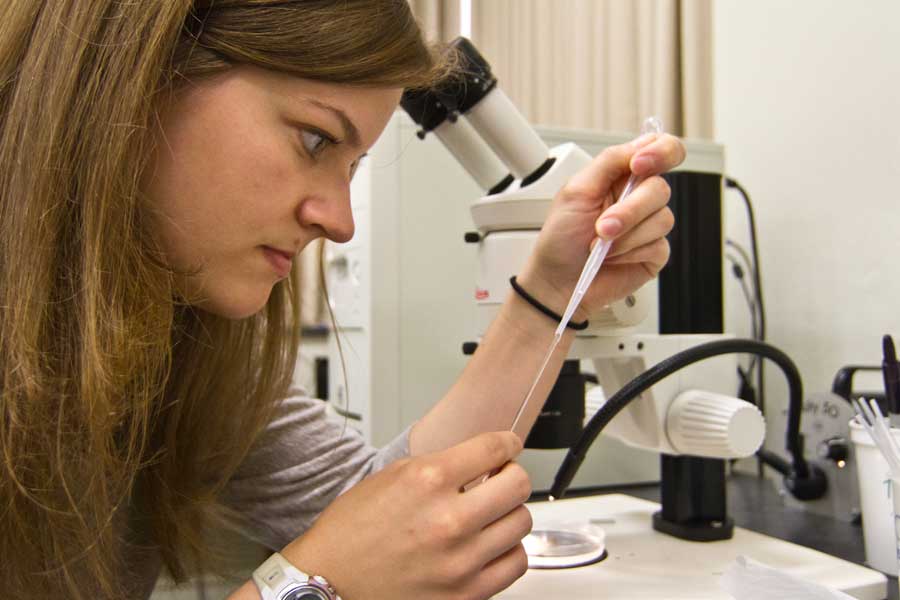
In collaboration with Professor Judy Thorn, Kelli Huebner's summer research involves imaging Xenopus laevis embryos in vivo throughout development using an ultra-compact MRI (UC-MRI). The research is funded through the National Institutes of Health (NIH), and Kelli's summer housing is supported by the Richter Scholarship. A native of Hanover Park, Illinois, Kelli is a senior neuroscience and chemistry major and Latin minor.
Tell us more about your research.
We are responsible for replicating the images from Lee et al. (2007). This includes imaging the early stages of development (cleavage), and also gastrulation (which we have already been able to successfully image). Then, I manipulate the raw images using ImageJ in order to bring out tissue contrast and be able to see everything possible within the embryo. It is very exciting to be able to see the endoderm, mesoderm, and ectoderm.
Describe your day-to-day experiences.
We usually run our experiment at the beginning of the week. This includes injecting a female frog with human chorionic gonadotropin (hCG) in order to make her ovulate. We then fertilize her eggs using testis and begin imaging a single embryo once we know that it is fertilized. If we get a successful embryo we could be imaging in the lab for 24 hours straight. If it is unsuccessful we usually wait until the next week to try again. Since we figured out the proper imaging parameters, it can be very disappointing when the fertilization is unsuccessful, but that's science.
How did you learn about this opportunity?
I was offered the opportunity to work on this project from Dr. Judy Thorn after I did research in her lab for part of last summer. I was originally hired to work in her lab cleaning dishes two years ago.
What has been the best part so far?
It took 24 hours, but the coolest part was when we got a normally dividing embryo and were able to see the different tissue layers moving as it was gastrulating.
Can you cite an example of how your experiences at Knox have helped you in your research?
Throughout my undergraduate in the sciences I have learned that what you expect to happen usually doesn't and that has helped me keep my patience as my weekly experiments are not always successful. They are not always successful because some embryos don't get fertilized -- some die, some divide abnormally -- and you can never predict what will happen with the single embryo that is chosen to image.
What inspired you to pursue the research?
Most imaging is done on fixed tissue and being able to image development in vivo is novel and very exciting to watch.
How do you think this research will benefit you?
This project has given me the opportunity to network with the man who built the machine, who is from Albuquerque, and also with researchers from The University of Iowa. The UC-MRI has showed me a different side of research; I have had to learn how the machine functions, which is mostly physics oriented and a different field than developmental biology.
Published on July 24, 2012

http://knox-fo-dss.ingeniuxondemand.com/news/summer-research-spotlight-kelli-huebner
Printed on Saturday, September 13, 2025